In this issue
Construction starts on new VDCPrice change for PCR testing
Changes to BVD earnotch pooled PCR testing
Antimicrobial susceptibility testing
Guarded swab kits improve sample collection
Abortion due to vaccination of young heifer with RB51
Pathology slide conference part of continuing ed activities
Vitosh passes first step toward certification
Leger to present at AAVLD annual meeting

Summer 2015
Construction starts on new VDC

Construction began July 1, 2015, on the new Veterinary Diagnostic Center. The new building, located northwest of the current VDC on UNL’s East Campus, will provide much needed space for laboratories, implementation of new technologies, new instrumentation and enhanced biosecurity.
For 38 years the current building has served the diagnostic needs of Nebraska’s animal industries. Over time space limitations forced some changes. When the VDC toxicologist retired, the decision was made to close that laboratory and use the space to expand the molecular diagnostic section, which was seeing an increase in testing demand. Today, the 9,083 square feet of useable space in the current building is inadequate, and several VDC faculty and support staff use laboratory space in another building. The new building will provide 40,582 square feet of assignable space.
It became clear that a new facility was needed when concerns over biosecurity of the building and a lack of space for expansion were noted in the 2006 and 2011 reviews by the Accreditation Committee for the American Association of Veterinary Laboratory Diagnosticians
With the help and collaboration of Nebraska’s legislature; the Nebraska Veterinary Medical Association; Nebraska’s livestock commodity groups, particularly the Nebraska Cattlemen’s Association; various biomedical, commercial, and pharmaceutical firms; the Office of the Vice-Chancellor of the Institute of Agriculture and Natural Resources; the University of Nebraska Foundation; and other allied groups, $46.2 million was pledged toward the construction.
RDG Planning and Design, an architectural firm in Omaha, Nebraska, completed plans for the new building in April, construction bids were solicited in May, and Sampson Construction Company, Lincoln, Nebraska received the construction contract with a bid almost $5 million under the initial estimate. Construction is expected to be completed by December 2016 with an occupation date of February 2017.
The VDC is grateful for the support it received from the numerous groups allied with Nebraska’s agricultural and animal industries. Many hours of discussion, planning, and coordination were donated by interested individuals who recognized the need for a new facility, understood its importance to the state, and were willing to invest their time and effort to see that it became a reality. Many thanks to each individual who supported and participated in this endeavor.
Price change for PCR testing
Due to the increased cost to run PCR testing, there will be an increase in the price for several of the pooled PCR tests beginning September 1.
This will still provide a significant cost savings over running PCR on each individual sample. Pool screening is built into the pricing, so if a Trichomonas foetus pool is detected to be positive, the individual samples will be tested at no charge (up to two pools per accession).
Changes to BVD earnotch pooled PCR testing
The VDC has recently changed the number of samples included in a pool for BVD PCR testing of earnotches from 30 to 48. Significant validation data indicate no sensitivity loss due to the increased number of samples. This change will result in significant cost savings to our clients.
For those who may be new to this testing, or as a reminder to those currently testing, please review the testing requirements below.
Please do not send earnotches for testing in plastic bags. Earnotches must be in tubes. Inexpensive tubes (25 cents) can be found at https://us.vwr.com/store, catalog number 60819-310. These are our preferred tubes for earnotches. Vacutainer (red top) tubes work as well.
Label all tubes with tube numbers. Please ship the tubes in numerical order (matching the order on your submission form) in blood tube boxes which can be obtained from the VDC for the cost of shipping only.
- Earnotches should be approximately 1 cm in length.
- To prevent cross contamination, the notcher should be disinfected with bleach then rinsed in water between each animal.
- Earnotches should be mailed as soon as possible and should be kept refrigerated until they are shipped on ice packs.
Animal IDs should be recorded on the submission paperwork or can be typed into an Excel file and emailed to vdc2@unl.edu.
Pricing for pools:
- 1-10 samples $32.00
- 11-15 samples $34.00
- 16-30 samples $59.00 ($1.96/sample)
- 31-48 samples $75.00 ($1.56/sample)
Samples within positive pools will be sorted out by immunohistochemistry for $4/sample.
If you have any questions prior to submitting earnotch samples, please call the lab, 402-472-1434.
Antimicrobial susceptibility testing
What is a breakpoint and how is it determined?
A breakpoint is the concentration of an antimicrobial agent above which an organism is considered to be resistant and below which it is considered susceptible and thus has a higher likelihood of successful treatment.
Several variables are used to establish breakpoints:
- In vitro activity and potency against each species of bacteria
- Achievable and sustainable serum drug concentrations
- Half-life
- Elimination
- Metabolism
- Toxicity
- Drug dosage
- Route of administration
Breakpoints are determined by the Clinical and Laboratory Standards Institute Subcommittee on Veterinary Antimicrobial Susceptibility Testing (CLSI-VAST) CLSI Vet-01. The VAST subcommittee is establishing new breakpoints for a variety of organism/species/site combinations.
To establish a breakpoint on a diagnostic report, several types of data are used, including in vitro susceptibility data from diagnostic laboratories. This can help determine MIC values that distinguish wild-type organisms from those that have acquired antibiotic resistance. These are called epidemiologic cut-off values or ECVs. Additionally phamcokinetic/phamacodynamic data are used to determine drug distribution in the patient along with clinical data from prospective efficacy trials. In prospective clinical trials, drug sponsors must test the antimicrobial drugs against the specific disease (organism/anatomic location) in a specific species to achieve a clinical breakpoint. Because this is very time consuming and costly many of the drug companies cannot conduct all of these trials for all possible veterinary applications, so many use breakpoints from human testing. This is why a drug may have a breakpoint with “NI”—no interpretation.
How to interpret susceptibility results?
S—Susceptible means the breakpoint is less than or equal to the breakpoint established by CLSI and that antimicrobial drug is recommended for treatment of that pathogen.
I—Intermediate means the breakpoint falls into a range where if the drugs are physiologically concentrated or a high dosage can be used, it may be used to treat that pathogen.
R—Means the breakpoint is greater than the breakpoint established by CLSI, and that antimicrobial drug is not recommended for the treatment of that pathogen.
NI—Means not interpretable. There are no breakpoints for that drug-bug-species-disease combination.
What is a minimal inhibitory concentration (MIC)?
An MIC is the lowest concentration of an antimicrobial agent that prevents growth of an organism in a susceptibility test. Each drug will have different concentrations based on the amount of drug tested, which is usually measured in µg/mL. For example, the drug concentration for ampicillin may be <=2 (S) and for Trimethoprim/Sulphamethoxazole <=256 (S) and both would be classified as susceptible. In the VDC, the broth microdilution test method is used, where each organism is grown in the presence of a liquid media containing a specific concentration of antimicrobial, incubated for a specified time, and then read to determine if growth was inhibited.
References
Blondeau, JM. 2007. STAT: Steps to antimicrobial therapy, antimicrobial resistance and mutant prevention concentration—a strategy for optimizing antimicrobial therapy for bovine respiratory disease. 1st ed.
Clinical Applications Antimicrobial Susceptibility Testing. 2010. Pfizer Animal Health.
Guarded swab kits improve sample collection
Bovine respiratory disease is an economically significant and complex disease caused by bacterial and viral pathogens. One challenge to diagnostic success is the collection of informative antemortem samples. Traditionally this has been done by swabbing nasal secretions from infected animals. However, research has shown that sampling of deep nasopharyngeal secretions, using guarded swabs, provides samples that are more representative of lung flora and the pathogens that may be present. Additionally, these swabs reduce the amount of contaminating environmental bacteria that collects in the external nares which can cause false negative testing results. To maximize your diagnostic testing, the VDC has assembled deep nasopharyngeal swab collection kits with instructions.
The kit includes:
- 33 inch double guarded swabs (Jorgensen or Continental)
- Amies transport media tubes (for bacterial culture)
- Virus transport media (for virus PCR)
- UNL VDC submission
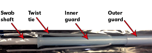
Sampling Protocol
- Sampled animals should be selected based on clinical signs and, ideally, in the acute phase of infection. If untreated animals are available, these should be selected first. Three to six representative animals should be selected for sampling.
- Restrain the head of the animal in a headgate in processing chute or other immobilization device.
- Remove dirt and debris from exterior nares with a clean cloth.
- Estimate the distance from the nares to the medial canthus of the eye.
- Unwrap the end of the swab with the twist tie and remove from the swab apparatus (this keeps the guards separate until ready for use).
- Insert the culture swab into the ventral meatus of the nose and advance it the estimated distance from the nostril to the medial canthus of the eye. Put slight pressure downward and toward the midline of the nose as you advance to ensure the swab stays in the ventral meatus and passes easily. Do not force the swab as this can cause excess bleeding and result in a poor sample.
- Retract the culture swab (not the guard) approximately 1-2 inches.
- Push the inner blue swab guard through the end of the outer guard.
- Push the swab through both guards and beyond the end of the inner guard for approximately 1-2 inches. Vigorously rotate the swab and advance and retract slightly (1/4”-1/2”) against the pharyngeal mucosa for 30-45 seconds. You want to ensure an adequate number of cells and exudate are collected in the sample.
- Retract the swab into the inner guard and then retract inner the guard into the outer guard.
- Remove the entire double guarded swab from the nares.
- Remove the inner guard containing the swab from the outer guard.
- Remove the swab from the inner guard.
- Using a clean pair of scissors, cut the cotton-tipped swab roughly 4-7 inches from the tip. To avoid contaminating the swab, do not let it touch any other surfaces. Do not cut the swab too short; short swabs are difficult to remove from the transport media.
- Open the sachet containing the amies transport media (Remel Bactiswab). You can remove and discard the swab with the blue cap included within the packet. Place the swab that you collected in the amies transport media and replace the white cap. Make sure the cotton-tipped swab is fully immersed in the gel.
- Repeat the procedure with a different double guarded culture swab in the other nostril. Place the swab in viral transport media and swirl vigorously. Make sure the tube cap is snapped firmly in place with two snaps or they may leak.
NOTE: swabs placed in virus transport media can’t be used for bacterial culture.
- Label all of the transport media legibly with the animal’s identification number or name.
- If the samples cannot be shipped immediately, they should be temporarily stored at 4° C in a cooler or refrigerator.
Shipping Requirements
- Fill out the VDC submission form with contact information and requested testing.
- For bacterial pathogens this usually consists of aerobic culture, antimicrobial sensitivity if required, and PCR for Mycoplasma bovis if suspected.
- Virology testing is the bovine respiratory disease multiplex PCR which includes BVD, IBR, BRSV and Bovine Coronavirus.
- Send the samples overnight with a sufficient number of ice packs to ensure they remain cold during shipment to the laboratory.
Please contact the bacteriology laboratory at 402-472-8470 if you have any questions.
Abortion due to vaccination of young heifer with RB51
By Dr. Bruce Brodersen
A 14-month-old heifer was bred while still with her dam. Not knowing she was pregnant, she was vaccinated against brucellosis on Jan. 15, 2015, using the RB51 vaccine strain of Brucella abortus. The fetus was expelled April 9, 2015. The attending veterinarian and an assistant were exposed to the fetus and fetal fluids. No gross lesions were reported in the fetus.
Among the tissues examined histologically, there was marked multifocal subacute necrotic and neutrophilic placentitis. Cytoplasm of numerous trophoblasts was distended by myriad coccobacilli. Bacterial cultures of placenta were positive for Brucella abortus. The isolate was confirmed as strain RB51 with PCR by the USDA-APHIS National Veterinary Services Laboratory.
Bovine brucellosis is a disease for which the USDA has implemented an eradication program due to the zoonotic potential of the disease. Vaccination plays a major role in the control strategy for brucellosis.
The most recently developed vaccine utilizes strain RB51. RB51 is a spontaneous rough mutant of B. abortus 2308 lacks the a homopolymer of perosamine as the O-chain component of LPS.1 The O-chain is an immunodominant antigen that can cause problems related to serologic diagnosis of vaccinated animals and is expressed in smooth colony types of B. abortus.1 In addition to lacking the O-side chain of LPS, this isolate is less virulent compared to known virulent strains and is protective against infection with virulent B. abortus, making it a suitable vaccine candidate.2
During infection, the majority of the organisms localize in cotyledons, placental membranes and in allantoic fluid. Early infection of the placenta begins in intercotyledonary erythrophagocytic trophoblasts followed by replication in adjacent chorioallantoic trophoblasts.3 After replication, there is necrosis of trophoblasts with release of massive numbers of organisms into the uterine lumen.3 The usual source of infection for cattle is ingestion of organisms from an aborted fetus or placenta, or contaminated uterine discharge.4
This case is unusual in that abortion was the result of administration of the vaccine strain (RB51) of B. abortus. It has been shown that vaccinating pregnant cattle with RB51 can result in rare placentitis and preterm expulsion of the fetus.5 Vaccination of pregnant cattle is ill-advised; therefore, vaccination is limited to young, non-pregnant cattle. In spite of recommendations, instances of fetal wastage occur.6 In the present case, the fact the pregnancy status was unknown resulted in fetal loss.
It should be noted that animal workers are at risk of infection by B. abortus when assisting parturition or handling infected tissues from these animals.
References
1 Schurig GG, Roop RM 2nd, Bagchi T, Boyle S, Buhrman D, Sriranganathan N. Biological properties of RB51; a stable rough strain of Brucella abortus. Vet Microbiol 28: 171-188, 1991
2 Poester FP, Goncalves VS, Paixao TA, Santos RL, Olsen SC, Schurig GG, Lage AP. Efficacy of strain RB51 vaccine in heifers against experimental brucellosis. Vaccine 24: 5327-5334, 2006
3 Anderson TD, Cheville NF, Meador VP. Pathogenesis of placentitis in the goat inoculated with Brucella abortus. II. Ultrastructural studies. Vet Pathol 23: 227-239, 1986
4 D.H. Schlafer RBM: Jubb, Kennedy and Palmer's Pathology of Domestic Animals, 5th ed., vol. 3, Saunders Elsevier, Philadelphia, PA, 2007
5 Palmer MV, Cheville NF, Jensen AE. Experimental infection of pregnant cattle with the vaccine candidate Brucella abortus strain RB51: pathologic, bacteriologic, and serologic findings. Vet Pathol 33: 682-691, 1996
6 Fluegel Dougherty AM, Cornish TE, O'Toole D, Boerger-Fields AM, Henderson OL, Mills KW. Abortion and premature birth in cattle following vaccination with Brucella abortus strain RB51. J Vet Diagn Invest 25: 630-635, 2013
Pathology slide conference part of continuing ed activities
By Dr. Bruce Brodersen
One of the continuing education activities that pathologists at the VDC take part in is the Wednesday Slide Conference (WSC) offered by the Joint Pathology Center (JPC) in Silver Spring, MD.
The WSC is a 25-week course where four cases are presented each week. Member institutions, including UNL, submit two cases for use in the course. In turn, the JPC redistributes case materials to the member institutions for use in their own training programs. These cases must have specific diagnoses and represent recognized, recently documented, or "classic" entities.
During the course the JPC sends histology slides to the institutions participating in the WSC, and the final reports are withheld until a later date, in order to challenge the participants. Pathologists and pathology residents review the cases during their respective weekly slide conferences and discuss the important features of the cases, disease mechanisms, and etiologic and morphologic diagnoses.
The JPC evolved from the joint entities at the Armed Forces Institute of Pathology. It is the federal government's pathology reference center supporting the Military Health System, Department of Defense and other federal agencies.
Vitosh passes first step toward certification

While the overall pass rate for all candidates who take the ACVP examination is approximately 33 percent, the pass rate for those who have completed the three year pathology residency program in the VDC is 100 percent.
Dr. Vitosh is the third individual to participate in the VDC's pathology residency program. She is also pursuing a PhD in the School of Veterinary Medicine and Biomedical Science. Her thesis topic is the study of the pathogenesis of porcine epidemic diarrhea virus and porcine delta-corona virus infections in swine.
In May Dr. Vitosh received an Othmer Fellowship, a highly coveted graduate student award given only to exceptionally qualified individuals who seek a terminal degree at UNL. Competition is university-wide and limited to only a few recipients each academic year.
Dr. Vitosh is a native of Odell, Nebraska and is a graduate of the ISU-UNL Professional Program in Veterinary Medicine. She was employed in a small animal practice in Beatrice, Nebraska prior to coming to UNL.
Leger to present at AAVLD annual meeting